You are here: BCU: Her2 Testing in Breast Cancer Management: Concordance of HER2 Status Between Primary and Metastatic Lesions  
Concordance of HER2 Status Between
Primary and Metastatic Lesions

Objectives
  • To compare HER2 overexpression and amplification in primary tumors and their distant metastases
  • To evaluate the HER2 status in different metastatic sites from the
    same patient.
Methods
  • 107 primary breast tumors and their corresponding distant metastases were analyzed by IHC, using the HercepTestTM, and by FISH.
  • HER2 status was also evaluated in 17 patients with at least 2 samples from metastatic lesions.
Results
  • The time between the removal of the primary tumor and the biopsy of the metastatic lesion ranged from one month to 18 years.

  • The discordance rate for HER2 overexpression between the primary and metastatic lesions was six percent. All six cases demonstrated greater HER2 overexpression in the metastatic lesion compared to the primary lesion.
  • The discordance rate for HER2 overexpression between different metastatic lesions was 18 percent.

  • The discordance rate with FISH between the primary and metastatic lesions was seven percent. Three cases were FISH-positive in the metastatic lesion but not in the primary lesion, and two cases were FISH-positive in the primary lesion but not in the metastatic lesion.

  • The discordance rate with FISH between different metastatic lesions was 19 percent.

Authors’ Conclusions
“This study does not support the routine testing of metastases to confirm HER-2 positivity when detected in the primary tumour, particularly if results obtained by FISH are available. Assessment of HER-2 status in one of the metastatic sites may be worthwhile only in some patients with easily accessible metastases and for whom HER-2 evaluation by IHC, recently performed in a primary tumour sample collected many years before, shows a negative score. An alternative solution would be the determination of HER-2 amplification by FISH in the primary tumour sample.”

Research Leader Commentary

The authors concluded that there’s no reason to retest metastases. For me, that would depend on the patient. If I had a patient with a HER2-negative primary tumor who had exhausted all avenues of treatment and wanted to try trastuzumab, I would try to biopsy a metastatic lesion to determine if it was positive because the tumor might respond to trastuzumab. Even if it were a 10 percent chance, I would take it in an individual patient who was motivated and was not responding to other therapies. Trastuzumab has relatively low toxicity and, in some cases, it has shown significant benefit.

Ann D Thor, MD

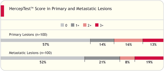
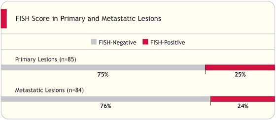
In our experience, it is highly unusual for the HER2 status to be altered during the development of the cancer. It is also very rare for us to find disagreement between the HER2 status of the invasive disease and the carcinoma in situ in the same patient. This is also true when we compare the primary tumor to the lymph-node metastasis.

In general, the HER2 status is quite similar or the same with only rare exceptions. In some of those exceptions, the morphologic appearance of the metastasis appears to be different, as if the tumor either developed new characteristics or was developed from an independent primary tumor.

Michael F Press, MD, PhD

Related Publications


Table of Contents Top of Page
Table of contents
 
CME Information
Editor’s Note:
Getting It Right
Faculty

Concordance Between Local and Central Laboratory HER2 Testing
- Related publications

Comparison of HER2 Assays
- Related publications
Concordance of HER2 Status Between Primary and Metastatic Lesions
- Related publications
HER2 Status and Response to Trastuzumab
- Related publications
College of American Pathologists
 - Related publications
 
Faculty Disclosures
Home · Contact us
Terms of use and general disclaimer